
Dedicated Ear & Hearing Care
Fellowship-trained care for complex ear, hearing, and skull base disorders
Hearing Loss
Types & Diagnosis
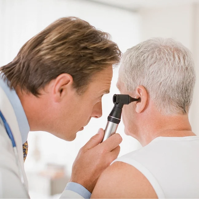
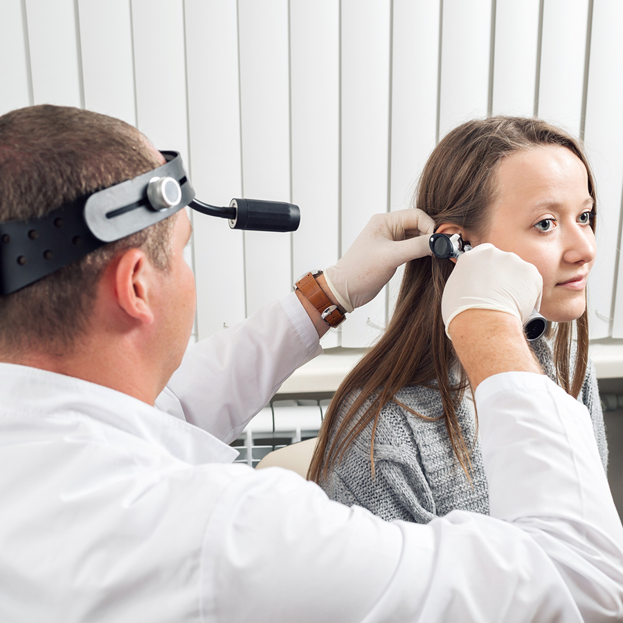
Hearing loss can affect one or both ears, appear suddenly or gradually, and range from mild to profound. It falls into three types: conductive (sound can't pass through the outer or middle ear, from causes like earwax, middle ear fluid, a torn eardrum, otosclerosis, or infection — often surgically fixable), sensorineural (inner ear or hearing nerve damage, from age, noise, or sudden hearing loss), or mixed (both). Diagnosis relies on a history, an ear exam, and hearing tests to find the type and any treatable cause.
Treatment & When to Get Checked
Treatment follows the cause: conductive issues (eardrum tears, ossicular problems, otosclerosis) often respond to surgery, while sensorineural loss may call for hearing aids or a cochlear implant evaluation. Get evaluated for any noticeable hearing change — especially sudden onset, one-sided loss, or hearing loss paired with tinnitus, dizziness, or ear fullness.
Sudden Hearing Loss
What It is & Why Speed Matters
Sudden sensorineural hearing loss (SSNHL) is a rapid drop in hearing — typically 30+ decibels within 72 hours — usually in one ear, sometimes with tinnitus, fullness, or dizziness. It's a medical emergency: treatment works best when started within the first few days, and delay worsens outcomes, so don't wait it out. Most cases are idiopathic (no clear cause), though viral infection, an inner ear vascular event, autoimmune disease, or (rarely) an acoustic neuroma can be factors; workup includes a hearing test and often an MRI.
Treatment & Outlook
First-line treatment is high-dose oral steroids; a steroid injection into the middle ear may be used first or as a backup if oral steroids don't work or aren't tolerated. Recovery varies — full, partial, or permanent loss are all possible — and earlier treatment improves the odds. Patients age 11+ are seen in Las Vegas and Henderson.

Eustachian Tube Dysfunction

What Causes the Blocked Feeling
The Eustachian tube connects the middle ear to the back of the nose and throat and balances ear pressure. When it doesn't open and close properly (ETD), pressure builds up — causing a blocked, full, or pressurized feeling without earwax buildup.
When Balloon Dilation May Help
For chronic ETD that hasn't improved with medical treatment — persistent pressure, fullness, muffled hearing, or trouble equalizing — Eustachian tube balloon dilation may be an option after a full evaluation confirms the diagnosis and fit.
Eustachian Tube Balloon Dilation
How the Procedure Works
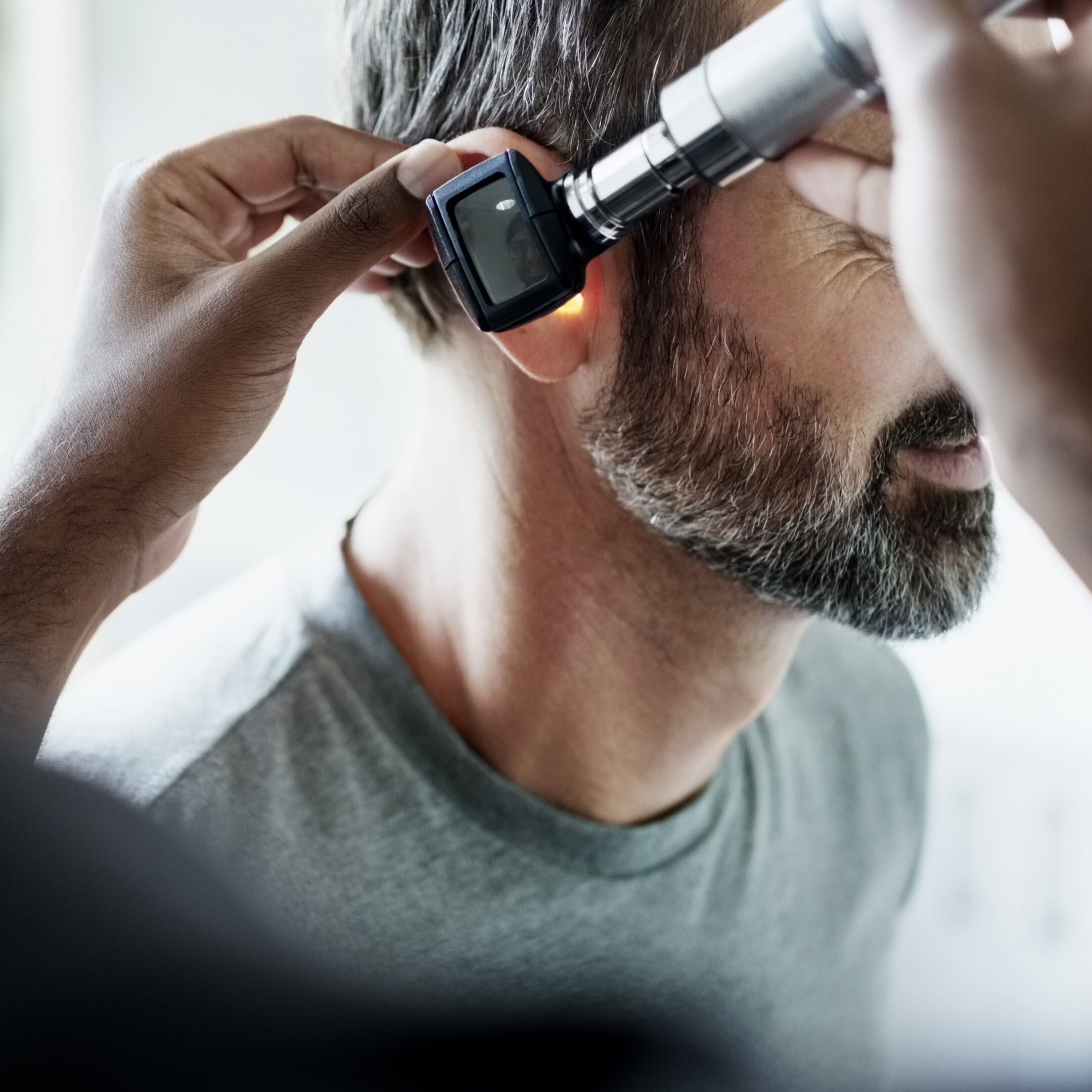
Eustachian tube balloon dilation is an in-office procedure for appropriate candidates with chronic ETD (persistent pressure, fullness, muffled hearing, or difficulty equalizing) that hasn't responded to medical treatment. A small balloon catheter is passed through the nose to the Eustachian tube opening, gently inflated to widen it, then removed — done under general anesthesia in just a few minutes per side.
Candidacy
Candidacy requires a thorough evaluation to confirm obstructive ETD and confirm the procedure is the right fit; Dr. Terrance J. Kwiatkowski performs this in Las Vegas and Henderson.
Chronic Ear Infections

Types of Infection
Ear infections (otitis media) are common; when they recur or don't clear, they can damage the eardrum and middle ear. Types include acute otitis media (sudden infection, usually after a cold, often self-resolving or antibiotic-treated), otitis media with effusion (middle ear fluid without active infection, causing muffled hearing for weeks or months), chronic suppurative otitis media (persistent/recurring drainage, often through a torn eardrum, that can cause hearing loss and complications if untreated), and otitis externa/"swimmer's ear" (ear canal infection, with recurring cases needing a look at underlying skin or anatomical causes).
Evaluation & Treatment
Evaluation includes an ear canal/eardrum exam, hearing tests, and a middle-ear function check, sometimes with imaging to rule out complications like cholesteatoma. Ear tubes may help recurrent infections or persistent fluid affecting hearing; tympanoplasty may be needed for chronic drainage or a torn eardrum, depending on the clinical picture.
Eardrum Perforation
Causes & Symptoms
A hole or tear in the eardrum can result from infection, trauma (cotton swab injury, a slap to the ear), a sudden pressure change, or as a side effect of ear tube placement; small tears often heal alone, larger or lasting ones usually don't. Symptoms include conductive hearing loss, drainage (especially with water exposure or infection), pain at the time of injury, tinnitus, and more frequent ear infections.
Repair & Recovery
Tympanoplasty repairs the perforation using a small graft (from tissue behind the ear or cartilage) to restore the eardrum, improve hearing, and reduce infection risk; damaged middle ear bones may be rebuilt at the same time or later (ossiculoplasty). Recovery involves keeping the ear dry for several weeks and avoiding pressure-raising activities, with hearing improvement typically noticeable within a few months as the graft heals.
Cholesteatoma
What It is & Symptoms
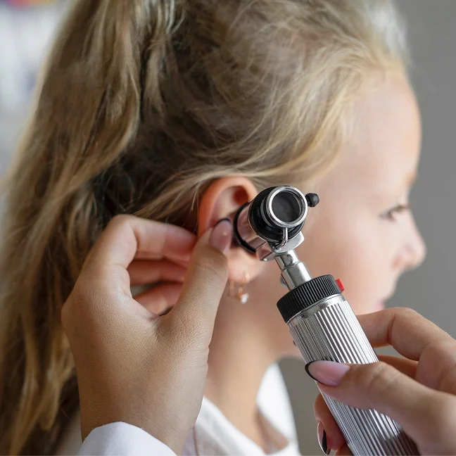
A cholesteatoma is an abnormal buildup of skin cells in the middle ear or mastoid — not a tumor, but still destructive if untreated, potentially eroding middle ear bones, harming hearing, injuring the facial nerve, or spreading to cause serious complications like meningitis or a brain abscess. It may be congenital (present from birth) or acquired — often from an eardrum retraction pocket linked to chronic ETD, or a torn eardrum edge. Symptoms include foul-smelling drainage, worsening hearing loss, ear fullness or pressure, dizziness (if the inner ear is involved), and facial weakness (a serious sign if the facial nerve is involved).
Surgical Treatment
Surgery is the only effective treatment, aiming for complete removal while protecting hearing and nearby structures — approaches include canal wall up or canal wall down mastoidectomy, sometimes with a second-look procedure to check for residual or returning disease. This is technically demanding, especially in revision cases or widespread disease, and draws on specialized neurotology fellowship training.
Otosclerosis/Stapes Surgery
What It is & Who's at Risk
Otosclerosis is abnormal bone growth in the middle ear, usually fixing the stapes (the body's smallest bone) in place, which blocks normal sound transmission and causes gradually worsening conductive hearing loss, typically starting in young adulthood. It often runs in families, is more common in women, usually begins in the 20s–30s, may worsen during pregnancy, and typically affects both ears (though not always equally). Symptoms include gradually worsening hearing (often noticed first in one ear), tinnitus, better hearing in noisy settings (paracusis Willisii), and sometimes dizziness or balance issues. Diagnosis uses audiometry (a Carhart notch can suggest otosclerosis), tympanometry, and CT imaging if unclear.
Surgical & Non-Surgical Options
Stapedectomy and stapedotomy surgically bypass or replace the fixed stapes with a small prosthesis to restore sound transmission and can be highly effective for the right patients; hearing aids are an alternative for those who prefer not to have surgery or aren't good candidates.
Mastoid Disease

How Infection Spreads to the Mastoid
The mastoid bone, located behind the ear, contains air-filled spaces connected to the middle ear, so infections there can spread into the mastoid — and conditions like cholesteatoma can too. Acute mastoiditis, a complication of acute otitis media, causes pain, swelling, and redness behind the ear and may need IV antibiotics and, if severe, surgical drainage. Chronic mastoid disease is most often linked to cholesteatoma or chronic suppurative otitis media, gradually damaging the mastoid and nearby structures.
Mastoidectomy
Mastoidectomy removes diseased tissue from the mastoid and is used for cholesteatoma, chronic mastoid infections, and related complications; the surgical approach (canal-wall-up or canal-wall-down) depends on disease extent and treatment goals. Mastoid procedures can be technically demanding, especially after a prior operation, and draw on specialized neurotology and skull base fellowship training.
Tinnitus
Causes
Tinnitus is the perception of sound (ringing, buzzing, hissing, clicking, roaring) without an outside source — a symptom, not a disease, that can affect one or both ears, be constant or intermittent, and range from mild to severely disruptive. Possible causes include noise- or age-related hearing loss, ETD or middle ear fluid, otosclerosis, eardrum perforation or middle ear disease, Ménière's disease, ototoxic medications, acoustic neuroma (rare but important to rule out), and cardiovascular conditions causing pulsatile tinnitus (a rhythmic sound following the pulse, which needs its own vascular workup).
Evaluation & Management
Evaluation includes a detailed history, otoscopy, and audiometry, with further testing/imaging for pulsatile, one-sided, or hearing-loss/dizziness-associated tinnitus. When a treatable cause exists, addressing it may help; otherwise, management includes sound therapy, cognitive behavioral therapy, or hearing aids — no medication is proven universally effective.
Dizziness & Vertigo

Inner Ear Causes
The inner ear's vestibular system signals the brain about head position and movement; disrupted signals cause vertigo (spinning), unsteadiness, or imbalance. Common inner ear causes: BPPV (the most common cause — displaced calcium crystals trigger brief, intense spinning with head movement, highly treatable with repositioning maneuvers), Ménière's disease (vertigo episodes with fluctuating hearing loss, tinnitus, and fullness, from abnormal inner ear fluid pressure), vestibular neuritis/labyrinthitis (viral infection causing sudden, severe vertigo that eases over days to weeks), and superior semicircular canal dehiscence (a bony opening causing dizziness triggered by sound or pressure).
Evaluation & Treatment
Evaluation includes a history, physical exam with vestibular testing, and audiometry, with VNG, VEMPs, or imaging as needed. Treatment depends on the cause: repositioning maneuvers for BPPV, diet/medication/procedures for refractory Ménière's, and vestibular rehab therapy for chronic vestibular dysfunction.
Ear Pressure/Barotrauma
Why It Happens & Signs
Ear pain, pressure, or fullness during or after flying is common, since rapid altitude changes can outpace the Eustachian tube's ability to equalize pressure; swallowing, yawning, or the Valsalva maneuver can help, but ETD may interfere. Barotrauma is injury from pressure differences, ranging from mild discomfort and temporary hearing loss to middle ear fluid, bleeding around the eardrum, or (in severe cases) a torn eardrum — diving carries more risk than flying due to faster, more pronounced pressure changes. Signs include ear pain/pressure during or after flying or diving, muffled hearing, lingering fullness, tinnitus, and (in severe cases) dizziness.
Management
Mild cases usually improve within hours to days; nasal decongestants and steroid sprays can help, but lasting hearing loss, dizziness, or drainage should be evaluated. When ETD drives recurrent barotrauma, treating it — including balloon dilation for appropriate patients — may help.

Complex & Revision Ear Surgery

When It's Needed
Revision ear surgery addresses an ear condition after a prior procedure didn't fully resolve it — often more challenging than initial surgery due to scar tissue, altered anatomy, and the reason the earlier procedure fell short. Reasons to consider it include cholesteatoma that persists or returns after mastoidectomy, hearing loss continuing after tympanoplasty or ossiculoplasty, ongoing drainage or middle ear disease after prior surgery or ear tubes, problems from a previous procedure, or wanting a second opinion before recommended surgery.
Specialized Training & Planning
Specialized neurotology and skull base fellowship training covers these complex cases — revision mastoidectomy, revision tympanoplasty, recurrent cholesteatoma, and complex ossicular reconstruction. Planning starts with reviewing prior operative reports, imaging, and hearing tests to understand the earlier procedure and why symptoms continued, guiding the next surgical approach.